
- 冠状ステント 冠状風船 カテーテル ガイドワイヤー イントロダクターシース アクセサリー 周辺介入制品
- 免疫アッセイ 分子診断 血液学診断 POCT ラボソリューション
- 構造心臓病製品
- 血管アクセス 血圧トランスデューサー 動脈血液サンプラー CVCアクセサリー IVカテーテル Needle-freeコネクタ インスリン注射 痛みの軽减
- 血液透析用消耗品 Hemoperfusionカートリッジ 透析マシン
- ECG 患者モニター オキシメータ
- 心臓血管 腫瘍学 抗感染薬 中枢神経系 その他
- 外科用ステープラー 内視鏡検査 内視鏡超音波システム
- トラウマインプラント 脊椎インプラント 人工ジョイント スポーツ医学
- ペースメーカーとリード 設備
- ELISA クラ FIA
- 核酸抽出 核酸抽出 & PCRキット リアルタイムPCR
- Thrombelastographyアナライザー 血液グループ化 血小板凝集
- POCT-FIA コロイド金 血糖 コレステロール SARS-CoV-2
- NeoECG Holterモニター ポータブルECGモニター AI-ECG ポケットECG
- 患者モニター バイタルサインモニター 潮間帯カプノグラフィー
- 指先オキシメータ ハンドヘルドOximeter 手首の酸化計 ウェアラブルOximeter オールインワンヘルスモニター POCTソリューション
- OBS ロック圧缩プレート ロックプレート 従来のプレート ミニプレートシステム ネジ Patellaリング-特許取得済み製品 金属ピン ケーブルシステム
- 内部固定システム Laminoplastyプレートシステム 経皮的カイフォプラスティ 融合ケージ 頸部固定システム
- ヒップシステム 膝システム
- 縫合アンカー 縫合ボタン
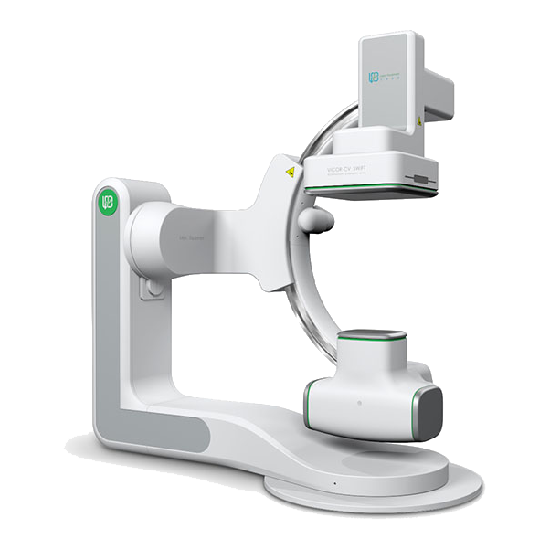
Vicor SWIFT医学の血管造影X線システムの特徴
1高い定义と安定したイメージ
正確で効率的な臨床診断をサポートする高解像度と広域画像
◆Digital flat panel detector
◆ Multiple panel models
◆ Adjustable, and wide-field
◆ Panel-digitized dynamic range up to 16 bit
◆ Accurate and clear details
◆ 解像度最大1956 × 1956
2 Long-lasting and Efficient Performance
◆3.0M heat capacity X-Ray tube, having long-lasting and stable life.
◆ Liquid-metal lubricated bearing to improve heat dissipation efficiency.
◆ トリプルフォーカススポット、さまざまな臨床アプリケーションに鮮明な画像を提供します。
3安全な放射線保護
◆ Accurate working of the collimator to effectively reduce scattered rays
◆ アクティブな放射線量の管理と監視、および放射線量の効果的な監督を実現するための運用中のRDSRによる放射線量とその他の情報のリアルタイムで正確な記録
◆ Function of virtual collimator, to locate the checked site without X-ray egression
◆ 複数の線量モデルでさまざまなニーズを満たし、低線量でも高品質の画像を取得
4プロのソフトウェアシステム
Vicor CV Workstation
◆ 64-bit operating system, providing user-friendly interactive interface
◆ Accelerated real-time processing module with efficient GPU, and stable and reliable image
◆ Supporting multiple image processing tools such as automatic measurement, ventricular analysis and QCA analysis
◆ Real-time function of virtual beam limiter, helpful to reduce scattered rays and improve the safety of both physicians and patients
◆ Radiation dose report in compliance with IEC standard
◆ ネットワーク安全基準に準拠した安全設計
Vicor AngioExpert
◆ DICOM3.0および関連する標準に準拠したデータ伝送
◆ Supporting data storage and migration management
◆ Efficient completion of vascular contour and morphological data analysis, and generation of QCA analysis report
◆ Supporting dynamic correction of stent position to enhance stent visualization
◆ 診断範囲を拡大し、手足の詳細を明確に表示するための全自動シームレス画像スプライシング
5すべての部署での臨床応用

6ステント強化
As an image recognition technique, it is used to realize the motion compensation for the X-ray image to clearly and accurately display the fine structure of the stent, and help physicians immediately evaluate the release of the stent during the operation.
* この機能には、販売に関する詳細な相談が必要です
7スウィフト3D
Vicor-CV SWIFTは、完璧な3D再構築機能を備えており、ボリューム再構築 (VR) 、多平面再構築 (MPR) 、最大強度投影 (MIP) 、最小強度投影 (MINIP) などの再構築手法をサポートしています。 血管の3次元形態と空間関係の分析に大きな利点があり、血管系疾患、腫瘍などの介入療法に大きな診断価値があります。
1.それは、血管の画像をより明確で正確にするために、血管構造の重複を避けることができます。
2.病変部位と他の正常組織および器官との間の3次元関係および卵管内腔構造は、任意の角度で観察することができる。
3.造影剤の投与量、診断と治療と手術の時間、および曝露量を減らすことができます。
4.正確な測定結果は、手術と介入治療のデータを提供できます
Lepuの医学のあなたの注意をありがとう!
質問やお問い合わせをメールで送信するか、連絡先データを使用してください。 私達はあなたに答えて幸せです 質問です。